- It all starts with the neuron.
- Cell body: the cells life-support center (nucleus) (also called soma)
- Dendrites: receive messages from other cells
- Axon: passed messages away from the cell body to other neurons, muscles, or glands
- Neural impulse: electrical signal traveling down the axon
- Myelin sheath: covers the axon of some neurons and helps speed neural impulse
- Terminal branches or axon
- NEURONS DO NOT TOUCH EACH OTHER THE SPACE IN BETWEEN IS CALLED THE SYNAPSE

How a neutron fires- it is an electrochemical process
- Electrical inside the neuron
- Chemical outside of the neuron (in the synapse in the form of a neurotransmitter)
- The firing is a call to action potential
All or none response-
- the idea that either the neuron fires or does not- no part way firing. Like a gun!

Steps of action potential
- Dendrites encorve neurotransmitters from another neuron across the synapses
- Reached its threshold, then fires based on the all or none response
- Opens a portal in the axon and lets positive ions (sodium) which mix with negative ions (potassium) that is already inside the axon (this neurons at rest have slightly negative charge.
- Mixing of + and - ions causes an electrical charge that opens up the next portal (letting in more k) while closing the original portal
- Process continues down axon to the axon terminal
- Terminal neurons turns electrical charge into chemical (neurotransmitter) and shoots message to the next neuron across the synapse

TYPES OF NEURONS
Sensory Neurons- take information from the senses to the brain. (Senses of touch and feel send messages to neurons. Any senses apply?)
Inter Neurons- take messages from sensory neurons to other parts of the brain or to motor neurons.
Motor Neuron (efferent neuron)- take information from the brain, to the rest of the body.
DRUGS CAN BE-


Parasympathetic nervous system:
THE BRAIN- 6 ways we study the brain
-Accidents: Phineas Gage story.... personality changed after accident. Told us that different part of
the brain control different aspects of who we are.
-Lesions: removal or destruction of some part of the brain. (Removing some part of the brain to study) frontal lobotomy.
-EEG: detects brain waves through their electrical output. Used paint in sleep research.
-CAT scan: 3D X-ray of the brain. Good for tumor locating but tell us nothing about function.
-PET scan: measures how much of a chemical the brain is using (usually glucose consumption)
-MRI: more detailed picture of the brain using magnetic field to knock electrons off axis. Takes many still pictures and turns images into a movie like a production.
-Functional MRI: combination of MRI and PET scan.

PARTS OF THE BRAIN. 🧠

Cerebral cortex: Madden up of densely packed neurons we call “gray matter”
Glial cells: support brain cells
-Wrinkles are called fissures
HEMISPHERES- two hemispheres.
- colateral control, right controls left and left controls right
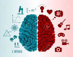
Right hemisphere ppl- special and creative tasks
Left hemisphere ppl- logic and sequential tasks
THE FOUR LOBES-

Sensory Neurons- take information from the senses to the brain. (Senses of touch and feel send messages to neurons. Any senses apply?)
Inter Neurons- take messages from sensory neurons to other parts of the brain or to motor neurons.
Motor Neuron (efferent neuron)- take information from the brain, to the rest of the body.
DRUGS CAN BE-
- Agonists- make neurons fire.
- Antagonismo- stops neurons from firing
- Reputarle inhibitors- block neurotransmitters from entering the neuron.
NERVOUS SYSTEM
- Central nervous system: the brain and spinal cord (CNS)
- Peripheral nervous system: all nerves that are not encased in bone (everything besides the cns) everything but the spinal chord (PNS). Divided into two categories, somatic and autonomic.
Somatic - uses motor (efferent neurons). Controls voluntary muscle movements.
Autonomic - controls the automatic functions of the body. Divided into two categories. The sympathetic and the parasympathetic.
Sympathetic nervous system:
- Fight or flight response
- Automatically accelerates heart rate, breathing, dilated pupils, slows down digestion
Parasympathetic nervous system:
- Automatically slows the body down after a stressful event.
- Heart rate and breathing slow down, pupils constrict and digestion spreads up.
REFLEXES- sensory neurons
ENDOCRINE SYSTEM- a system of glands that secrete hormones. Similar to nervous system, except hormones work a lot slower than neurotransmitters.
THE BRAIN- 6 ways we study the brain
-Accidents: Phineas Gage story.... personality changed after accident. Told us that different part of
the brain control different aspects of who we are.
-Lesions: removal or destruction of some part of the brain. (Removing some part of the brain to study) frontal lobotomy.
-EEG: detects brain waves through their electrical output. Used paint in sleep research.
-CAT scan: 3D X-ray of the brain. Good for tumor locating but tell us nothing about function.
-PET scan: measures how much of a chemical the brain is using (usually glucose consumption)
-MRI: more detailed picture of the brain using magnetic field to knock electrons off axis. Takes many still pictures and turns images into a movie like a production.
-Functional MRI: combination of MRI and PET scan.
PARTS OF THE BRAIN. 🧠
- Hind- controls basic biological structures.
- Medulla- located above spinal chord. It is involved in the control of breathing, heart rate, and blood pressure.
- Pons- located above the medulla conecta the hind brain, mid brain and forebrain. It is involved in facial expressions (emotion)
- Cerebellum- bottom tear of the brain. Known as the little brain. Coordinated fine muscle movements.
- Mid brain- coordinates simple movements with sensory information. The most important structure in the mid brain is the reticular formation. (Controls arousal and the ability to focus our attention)
- Fore brain- makes us human. Largest part of the brain. Made up of the thalamus, the limbic system, and the cerebral cortex.
- Thalamus- switchboard of the brain. Receives sensory signals from the spinal chord and sends them to other parts of the forebrain. Every sense except small 👃
- Limbic system - hypothalamus, pituitary gland, amygdala, and hippocampus.
- hypothalamus: the most important structure of the brain. Controls and regulates body temperature, sexual arousal, hunger, thirst, and the endocrine system.
- Amygdala- involved in how we process memory. And deals with emotions.
- hippocampus- involved in the processing and storage of memories.
Glial cells: support brain cells
-Wrinkles are called fissures
HEMISPHERES- two hemispheres.
- colateral control, right controls left and left controls right
Right hemisphere ppl- special and creative tasks
Left hemisphere ppl- logic and sequential tasks
THE FOUR LOBES-
- Frontal lobes: thought and emotional control. Contains motor cortex. And sends signals to tour body controlling muscle movement. Contains Broca’s area which is responsible for controlling muscles that produce speech. Damage to Broca’s area is called Broca’s aphasia- unable to make movements to talk.
- Parietal lobe- contains the sensory cortex which receives incoming touch sensations from rest of the body most of the parietal lobe is made up of association areas. Association area- any area not associated with receiving sensory information or coordinating muscle movements.
- Occipital lobes: deals with vision. Contains visual cortex which interprets messages from our eyes into images we can understand.
- Temporal lobe: deals with hearing. Sound processed through the ears! Interpreted in the auditory cortex. Contains Wernikie’ s area- interprets written and spoken speech. Wernickies aphasia- unable to understand language. The syntax and grammar is jumbled.
Brain plasticity- the idea that the brain, when damaged, will attempt to find new ways to reroute
messages. Children’s brains are more plastic than adults.
Corpus Callosum- thick bundle of nerves.

The organization is really good ,as well as, it's very visually pleasing. I feel like I could study and understand this very well. I can see where everything is, how its categorized, and the explanation of everything. Good job!
ReplyDelete